Clinical Results & Findings
Proven results treating central airway obstruction.
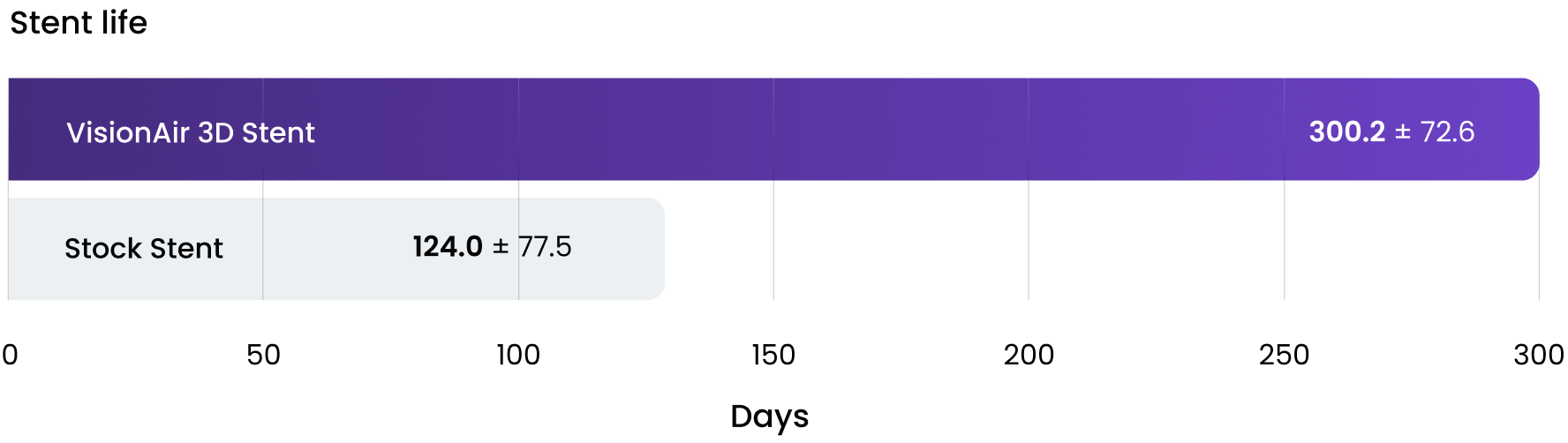
VisionAir 3D Stents provide a longer stent life than standard stock airway stents, resulting in an increased time between stent replacements.
How are physicians using VisionAir 3D Stents?
VisionAir 3D Stents currently treat a variety of underlying diagnoses including complex disease where patients present with multiple types of obstruction.
Complex vs. Single Disease

Current Use Statistics

*Data current as of 2023
Our stents have broad FDA-cleared indications for patients with symptoms associated with central airway obstruction (CAO).
VisionAir 3D Stents last longer than traditional stock stents.
VisionAir 3D Stents provide a longer stent life than traditional stock airway stents resulting in an increased time between procedures.


Proven results treating Central Airway Obstruction (CAO).
VisionAir 3D Stents provide a longer stent life than standard stock airway stents, resulting in an increased time between stent replacements & procedures.

Proven results treating Central Airway Obstruction (CAO)

Clinical Use Cases


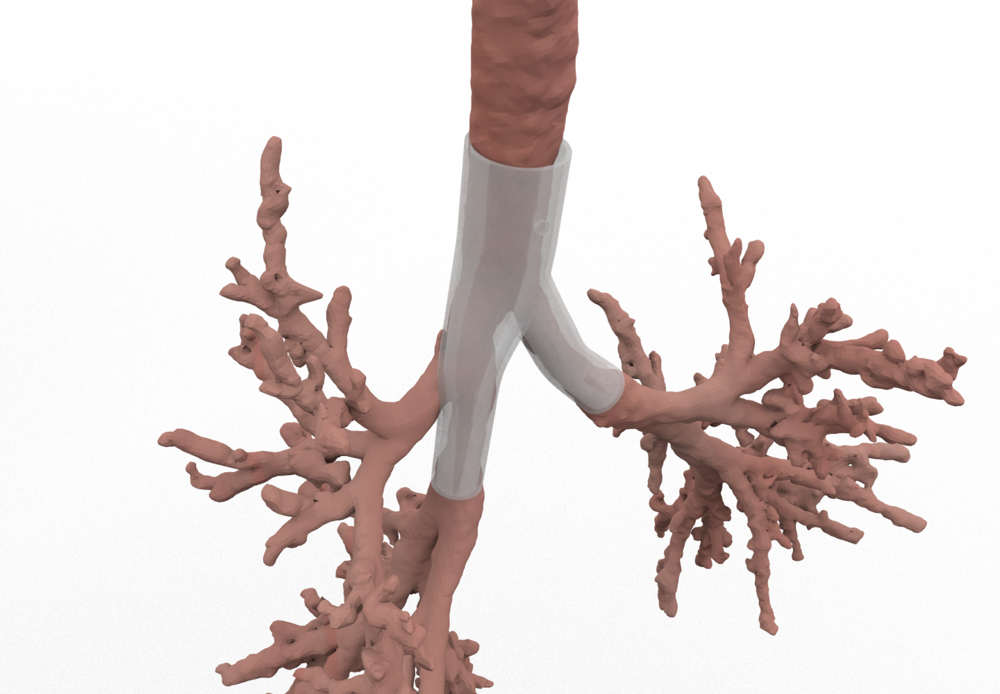

Tracheobronchomalacia (TBM)
The length of the 3D stent was based off the malacia at the thoracic inlet and extended distally into the left main stem. Sizing was a challenge due to the shape of the airway being a flattened ellipse, therefore, estimating the diameter was determined using the calculated long and short axes of the airway. After the main carina, variable sizing in diameter was required due to the significantly larger right main stem compared to the left main stem.

Tracheobronchomalacia (TBM)
The left main stem was sized so that the stent would directly interact with the luminal wall of the airway, using the 1 mm wall thickness to provide support. The stent was designed to contour the luminal path of the airway in order to prevent misalignment or excessive pressure on the airway wall of the upper lobe, where the airway stent branch extended past into the bronchus intermedius.
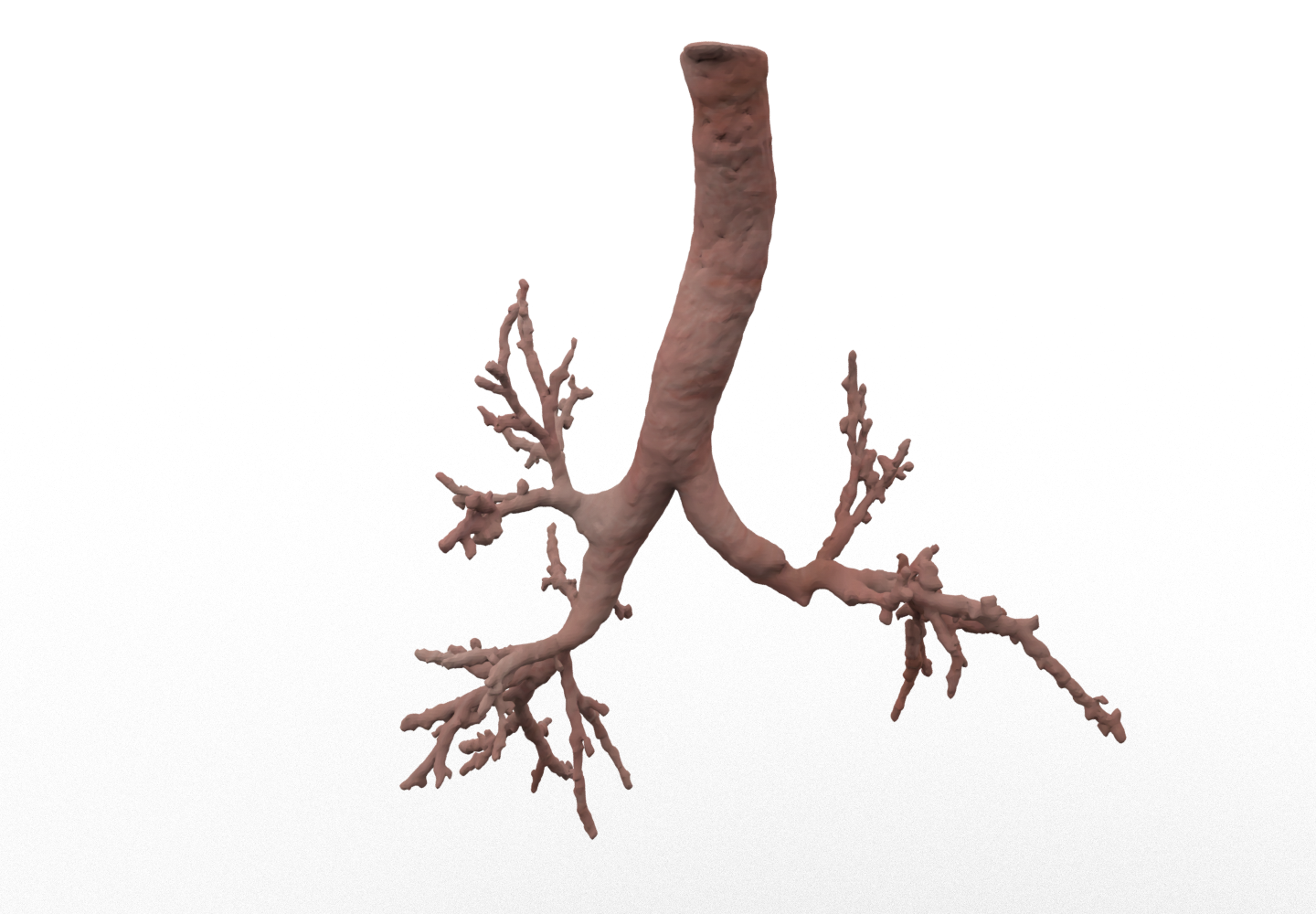
Bronchomegaly
Patient presented with bronchomegaly. The patient had focal right-sided malacia with recurrent infection. Stenting of the right main branch over the years with stock stents were purposely oversized with the intention to prevent migration but had appeared to cause dilation of the native airway.


